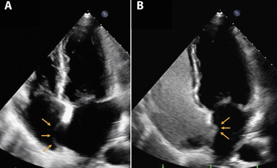
You may have already had an echocardiogram (sometimes just called ‘echo’) performed. This is a non-invasive imaging test using ultrasound to look at your heart. Ultrasound is very high frequency sound which cannot be heard by the human ear. It is used to gain information regarding the structure and function of the heart muscles, chambers of the heart and structures within the heart such as the valves. The test is painless and does not use radioactivity. A bubble contrast echocardiogram uses imaging ultrasound combined with an injection of micro bubble contrast to help determine additional information.
Your doctor suspects that you may have a hole in your heart. The majority of significant holes in your heart are detected in childhood. However, if there is a small defect or hole in the wall (inter-atrial septum) separating the left and right upper chambers of the heart (atria), this may not come to light until adulthood. The micro bubble contrast allows for the detection of these small holes as they do not usually show up on a normal echocardiogram.
It also helps in diagnosing Pulmonary AV Fistulas in patients with End Stage Liver Disease who are undergoing liver transplant to anticipate difficulties in ventilation during the surgery.
